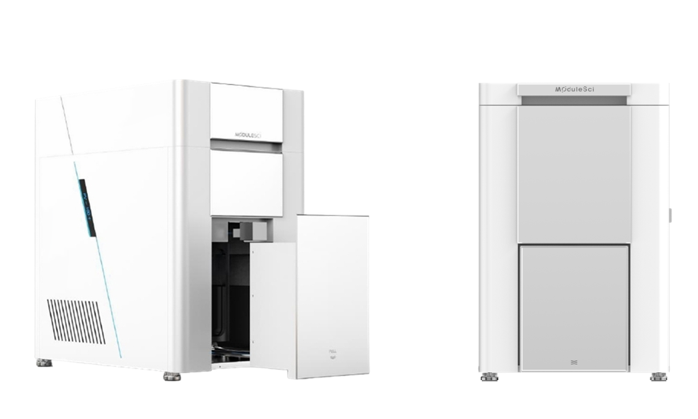
Key Features
- Auto-extendable BSE detector: a retractable four-segment backscattered-electron detector with high-sensitivity semiconductor elements optimizes SE imaging at short working distances and gives extra clearance for large, tall or complex-shaped specimens, minimizing collision risk
- Compact benchtop design with no complex installation and a minimal footprint, run from an intelligent, easy-to-learn user interface
- Resolution of 5 nm at 30 kV, with magnification from 30× to 300,000×
- Low vacuum mode observes non-conductive samples — ceramics, plastics, fibers, biological tissue — directly, without gold or carbon sputter coating
- Variable pressure mode from 10–150 Pa in 10 Pa steps, with gas-assisted charge compensation that suppresses charging artifacts, image drift and brightness fluctuation
- Optical navigation with one-button automatic brightness/contrast, focus and astigmatism adjustment
- Optional SE/BSE/EDS detection and image-stitching modules
Specifications
| Parameter | Specification |
|---|---|
| Emission source | Pre-aligned tungsten filament electron gun |
| Resolution | 5.0 nm |
| Magnification (photo) | 30× ~ 300,000× |
| Accelerating voltage | 1~30 kV |
| Detector | Secondary Electron Detector (ETD); optional Backscattered Electron Detector (BSED), EDS |
| Image format | TIFF, JPG, BMP, PNG |
| High vacuum mode | ≤5×10⁻⁴ Pa |
| Low vacuum mode (optional) | 6~150 Pa |
| Automatic stage | 3-axis (5-axis optional) |
| Stage moving range | X: 0~50 mm, Y: 0~50 mm, Z: 4~35 mm |
| Main unit dimensions | 400(W) × 620(D) × 680(H) mm |
| Weight | 110 kg |
| Room requirements | 2m × 2m × 2m |
| Power supply | AC 110/220V (±10%), 50/60Hz, 1.5kW |
| Temperature & humidity | 20±5°C, ≤60%RH |
Demonstrated on fiber samples at 20 Pa, 60 Pa and 150 Pa, the variable-pressure system holds stable, distortion-free imaging across the full range — with no gold or carbon coating required. A higher-throughput floor-standing sibling (PE-100) and a field-emission model (PE-300) are also available in the ModuleSci SEM range.
Intelligent Operating Software
ModuleSci’s self-developed SEM software captures an image in three guided steps: mount and set the sample, take a navigation image while the chamber evacuates, then navigate to the region of interest and capture the SEM image. Automatic brightness/contrast, focus and astigmatism adjustment run in the background, with live chamber monitoring throughout.
Photo-assist tools include dimension measurement, image mixing and gesture-based quick navigation, with optional image stitching for wide-field views.
Software at a Glance
- Workflow
- Mount sample → navigate & evacuate → locate ROI & capture
- Languages
- Korean, English, Chinese, French, German, Russian
- Photo-assist tools
- Dimension measurement, image mixing, gesture navigation, optional image stitching
About the Tabletop SEM
The PicoVision-100P (PV-100P), also called the Table-top W-SEM, is a compact tabletop scanning electron microscope from ModuleSci, South Korea, with a tungsten-filament electron gun, 5 nm resolution at 30 kV and magnification from 30× to 300,000×. Its retractable four-segment backscattered-electron detector gives extra clearance for large, tall or complex-shaped specimens.
Low vacuum and variable pressure modes let it image non-conductive samples such as ceramics, plastics, fibers and biological tissue without gold or carbon sputter coating. The variable pressure mode runs from 10 to 150 Pa in 10 Pa steps with gas-assisted charge compensation, which suppresses charging artifacts, image drift and brightness fluctuation. Optical navigation with one-button automatic brightness/contrast, focus and astigmatism adjustment keeps operation simple, and the compact benchtop design needs no complex installation.
Tabletop SEM: frequently asked questions
What are the resolution and magnification of the PicoVision-100P?
5.0 nm at 30 kV, with a photo magnification of 30× to 300,000× and an accelerating voltage of 1–30 kV, from a pre-aligned tungsten filament electron gun.
Can the PV-100P image non-conductive samples without coating?
Yes. Low vacuum mode observes non-conductive samples, such as ceramics, plastics, fibers and biological tissue, directly, without gold or carbon sputter coating. The variable pressure mode runs from 10 to 150 Pa in 10 Pa steps with gas-assisted charge compensation, which suppresses charging artifacts, image drift and brightness fluctuation.
Which detectors and options are available?
A secondary electron detector (ETD) is standard. A backscattered electron detector (BSED), EDS and an image-stitching module are optional. The retractable four-segment BSE detector gives extra clearance for large, tall or complex-shaped specimens and minimizes collision risk.
What are the size and installation requirements of the PV-100P?
The main unit measures 400 × 620 × 680 mm (W × D × H) and weighs 110 kg. The room should be 2 m × 2 m × 2 m at 20 ± 5 °C and ≤60% RH, with AC 110/220 V (±10%), 50/60 Hz, 1.5 kW. The stage is 3-axis, with 5-axis optional.
Where can I buy the ModuleSci Tabletop SEM in India?
Multiflo Instruments, based in Navi Mumbai, supplies the ModuleSci SEM range in India. Our team can advise on the PV-100P, send a quotation, and arrange installation, training, calibration and after-sales service. Use the Contact Us button or call +91-22-44516175.
Related electron microscopy and analysis
Floor SEM
ModuleSci PicoEye-100 compact floor-standing SEM
Field Emission SEM
ModuleSci PicoEye-300 Schottky FESEM, 0.9 nm
X-Ray Diffractometer
Linev Systems Tellus 300/600 benchtop XRD
Benchtop WDXRF
Spectron MAKC GVM, sodium to uranium in one measurement
Plasma Cleaners
Digit Concept iSP 3 plasma cleaner
Plasma Decapsulator
Digit Concept iMIP-Dcap II for IC failure analysis
Get A Free Instrument Consultation!
We can suggest products to our customers to make them the leaders in their respective industries.