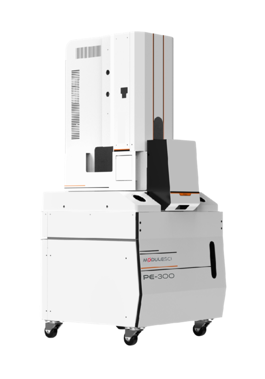
Key Features
- Advanced Schottky-type field emission electron source paired with a high-performance electron optical system for ultra-high imaging resolution
- Resolution of 0.9 nm at 15 kV and 1.3 nm at 1 kV, with high-quality imaging at both conventional and low accelerating voltages
- Low-voltage imaging observes non-conductive specimens directly, without conductive coating, preserving true surface morphology
- Integrates SE imaging, BSE imaging and EDS on a single platform for comprehensive morphology, microstructure and elemental characterization
- Rapid image acquisition with a large depth of field
- Large-travel sample stage and an In-lens Detector (ILD) alongside the standard secondary-electron detector
- Suited to research institutes, university laboratories and industrial quality control
Specifications
| Parameter | Specification |
|---|---|
| Emission source | Schottky-type thermal field emission electron source (ZrO/W) |
| Resolution | 0.9 nm @ 15kV, 1.3 nm @ 1kV |
| Magnification (photo) | 8× ~ 2,000,000× |
| Accelerating voltage | 0.02~30 kV |
| Detector | Secondary Electron Detector (ETD), In-lens Detector (ILD); optional Backscattered Electron Detector (BSED), EDS |
| Image format | TIFF, JPG, BMP, PNG |
| High vacuum mode | ≤2×10⁻⁵ Pa |
| Automatic stage | 5-axis |
| Stage moving range | X: 0~130 mm, Y: 0~130 mm, Z: 2~50 mm |
| Main unit dimensions | 1000(W) × 1500(D) × 1700(H) mm |
| Weight | 800 kg |
| Room requirements | 4m × 4m × 2m |
| Power supply | AC 110/220V (±10%), 50/60Hz, 4kW |
| Temperature & humidity | 21±4°C, ≤60%RH |
As a field-emission system, the PE-300 runs high vacuum only and uses low-voltage imaging — rather than a variable-pressure mode — to image non-conductive samples without coating. A tabletop model (PV-100P) and a compact floor-standing tungsten SEM (PE-100) are also available in the ModuleSci SEM range.
Application Examples
A sample of imaging results produced on the PE-300.
Porous Hollow SiO2 Spheres
5kV, SE imaging
Chitosan
20kV, SE imaging
RF Microsphere Aggregates
20kV, SE imaging
ZIF-8
20kV, SE imaging
Carbonized RF Spheres
20kV, SE imaging
Grain Particle Reference Standard
20kV, SE imaging
Intelligent Operating Software
ModuleSci’s self-developed SEM software captures an image in three guided steps: mount and set the sample, take a navigation image while the chamber evacuates, then navigate to the region of interest and capture the SEM image. Automatic brightness/contrast, focus and astigmatism adjustment run in the background, with live chamber monitoring throughout.
Photo-assist tools include dimension measurement, image mixing and gesture-based quick navigation, with optional image stitching for wide-field views.
Software at a Glance
- Workflow
- Mount sample → navigate & evacuate → locate ROI & capture
- Languages
- Korean, English, Chinese, French, German, Russian
- Photo-assist tools
- Dimension measurement, image mixing, gesture navigation, optional image stitching
About the Field Emission SEM
The Field Emission SEM (model PicoEye-300, PE-300) from ModuleSci, South Korea, pairs a Schottky-type field emission electron source with a high-performance electron optical system for ultra-high imaging resolution: 0.9 nm at 15 kV and 1.3 nm at 1 kV. Magnification runs from 8× to 2,000,000× and the accelerating voltage from 0.02 to 30 kV.
Low-voltage imaging lets the PE-300 observe non-conductive specimens directly, without conductive coating, preserving true surface morphology. SE imaging, BSE imaging and EDS can be combined on a single platform for morphology, microstructure and elemental characterization, with a large-travel 5-axis stage and an in-lens detector (ILD) alongside the standard secondary-electron detector. It suits research institutes, university laboratories and industrial quality control.
Field emission SEM: frequently asked questions
What resolution does the PicoEye-300 FESEM achieve?
0.9 nm at 15 kV and 1.3 nm at 1 kV, with a photo magnification of 8× to 2,000,000× and an accelerating voltage of 0.02–30 kV.
Can the PE-300 image non-conductive samples without coating?
Yes, by low-voltage imaging. As a field-emission system the PE-300 runs high vacuum only and does not use a variable-pressure mode; low-voltage imaging observes non-conductive specimens directly, without conductive coating, preserving true surface morphology.
Which detectors does the PE-300 support?
A secondary electron detector (ETD) and an in-lens detector (ILD) are standard. A backscattered electron detector (BSED) and EDS are optional. Images are saved as TIFF, JPG, BMP or PNG.
What are the installation requirements of the PE-300?
The main unit measures 1000 × 1500 × 1700 mm (W × D × H) and weighs 800 kg. The room should be 4 m × 4 m × 2 m at 21 ± 4 °C and ≤60% RH, with AC 110/220 V (±10%), 50/60 Hz, 4 kW. High vacuum is ≤2×10⁻⁵ Pa.
Where can I buy the ModuleSci FESEM in India?
Multiflo Instruments, based in Navi Mumbai, supplies the ModuleSci SEM range in India. Our team can advise on the PE-300, send a quotation, and arrange installation, training, calibration and after-sales service. Use the Contact Us button or call +91-22-44516175.
Related electron microscopy and analysis
Benchtop SEM
ModuleSci PicoVision-100P tabletop SEM
Floor SEM
ModuleSci PicoEye-100 compact floor-standing SEM
X-Ray Diffractometer
Linev Systems Tellus 300/600 benchtop XRD
Benchtop WDXRF
Spectron MAKC GVM, sodium to uranium in one measurement
Plasma Cleaners
Digit Concept iSP 3 plasma cleaner
Plasma Decapsulator
Digit Concept iMIP-Dcap II for IC failure analysis
Get A Free Instrument Consultation!
We can suggest products to our customers to make them the leaders in their respective industries.